This instructional guide provides a comprehensive overview of the female reproductive system using a model. We'll explore each component, its function, and how to effectively utilize the model for educational purposes. Understanding this system is crucial for reproductive health awareness, and this guide aims to make that understanding accessible and engaging. For additional resources, check out this helpful female reproductive model.
Model Overview: A Closer Look
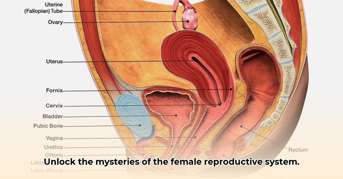
Your female reproductive system model is a three-dimensional representation of the organs involved in reproduction. It typically includes the following components, each carefully scaled and positioned to reflect their actual anatomical relationship:
- Ovaries (ov-a-reez): These almond-shaped organs are responsible for producing eggs (ova) and hormones like estrogen and progesterone (essential for menstruation and pregnancy).
- Fallopian Tubes (fal-lo-pee-an tubes): These slender tubes connect the ovaries to the uterus. They are the site of fertilization, where the sperm meets the egg.
- Uterus (yoo-ter-us): This pear-shaped, muscular organ is where a fertilized egg implants and develops into a fetus.
- Cervix (サービックス): The lower, narrow part of the uterus, connecting the uterus to the vagina. It dilates during childbirth.
- Vagina (va-jy-na): The muscular canal that extends from the cervix to the external genitalia. It serves as the birth canal and the passageway for menstrual flow.
Many models also include ligaments (supporting structures) and blood vessels to provide a more complete anatomical picture. Examine the model carefully, noting the relative size and positioning of each component. How does the spatial arrangement facilitate the process of reproduction?
Component Functions: A Detailed Exploration
Each component plays a vital role in the complex process of reproduction:
Ovaries: These organs release mature eggs (ova) in a cyclical process called ovulation. This process is regulated by hormones, and the released eggs travel down the fallopian tubes.
Fallopian Tubes: These tubes transport the egg from the ovary to the uterus. Fertilization typically occurs within the fallopian tubes, where sperm encounter and fertilize the egg.
Uterus: The uterus is the site of embryo and fetal development. Its lining thickens during the menstrual cycle in preparation for potential pregnancy. If fertilization doesn't occur, the lining is shed, resulting in menstruation.
Cervix: The cervix acts as a protective barrier between the uterus and the external environment. During labor, it dilates to allow the passage of the baby.
Vagina: The vagina serves as the birth canal during childbirth and the passageway for menstrual blood. It also plays a crucial role in sexual intercourse.
Did you know that the average menstrual cycle lasts about 28 days? This highlights the remarkable cyclical nature of the female reproductive system.
How to Use the Model: A Step-by-Step Guide
Initial Inspection: Carefully unpack your model and identify each component. Familiarize yourself with the provided labels and diagrams.
Spatial Relationships: Observe the relative positions of the ovaries, fallopian tubes, uterus, cervix, and vagina. How are these organs positioned to facilitate the journey of the egg and potential fertilization?
Visualizing the Process: Trace the path of an egg from the ovary, through the fallopian tube, to the uterus. Visualize the processes of ovulation, fertilization, implantation (if fertilization occurs), and menstruation. This hands-on approach significantly enhances understanding.
Interactive Learning: Explain the functions of each component to a friend or family member. The act of teaching reinforces your own understanding. This interactive learning is a key benefit of using a physical model.
Advanced Simulation: Some models allow for the simulation of processes like fertilization and implantation. If your model includes such features, utilize them to further deepen your understanding.
According to Dr. Evelyn Reed, Professor of Obstetrics and Gynecology at Harvard Medical School, "Using a physical model provides a concrete and engaging way to grasp the intricate workings of the female reproductive system. It transforms abstract concepts into tangible experiences."
Troubleshooting: Addressing Common Issues
Loose Parts: If any parts feel loose, ensure they are correctly aligned and securely fastened. Refer to the manufacturer's instructions for proper assembly.
Damaged Parts: Contact the manufacturer or retailer for replacement parts if any components are damaged.
Unclear Instructions: Consult online resources such as reputable medical websites or educational videos for further assistance.
Conclusion: The Value of Visual Learning
The female reproductive system is a complex and fascinating system. Using a physical model provides a valuable visual aid, translating complex anatomical concepts into a tangible learning experience. By actively engaging with the model, you gain a deeper and more intuitive understanding of this vital system. Remember, continuous exploration and learning are key to appreciating the wonder of the female reproductive system.